
Introduction: The Promise of Bioactive Glass in Regenerative Medicine
In the modern pursuit of bone regeneration and tissue engineering, the combination of stem cells with bioactive materials has emerged as one of the most promising strategies. Among these, bioactive glass has become a material of profound interest due to its unique ability to stimulate bone formation, enhance cell signaling, and promote biomineralization.
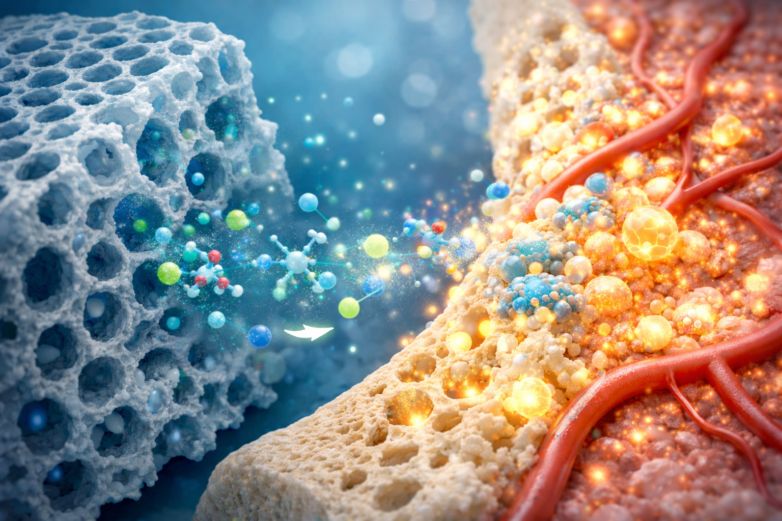
Bioactive glass serves not only as a structural scaffold but also as a biochemical messenger, influencing cellular behavior through the release of ions such as calcium, phosphate, and silicon. These ions activate crucial pathways in stem cells, guiding them to differentiate into osteoblasts — the bone-forming cells that are central to skeletal repair and regeneration.
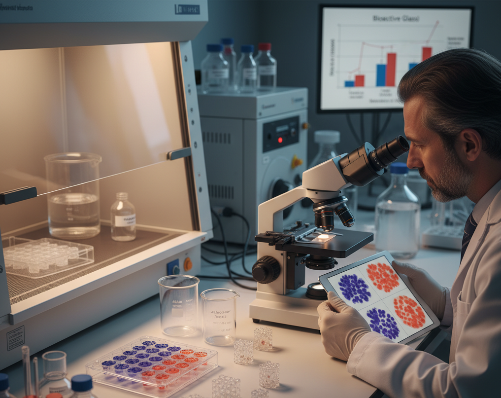
To validate and measure the osteogenic potential of bioactive glass, researchers rely on staining and assay techniques that provide insight into stem cell differentiation stages. Two key methods stand out:
- Alkaline Phosphatase (ALP) Assay – an indicator of early osteogenic differentiation.
- Alizarin Red S (ARS) Staining – a marker of late-stage mineralization and calcium deposition.
Both techniques are critical for understanding how bioactive glass enhances osteogenesis, enabling scientists to optimize formulations and applications for clinical and biomedical use.
Understanding the Role of Bioactive Glass in Bone Regeneration
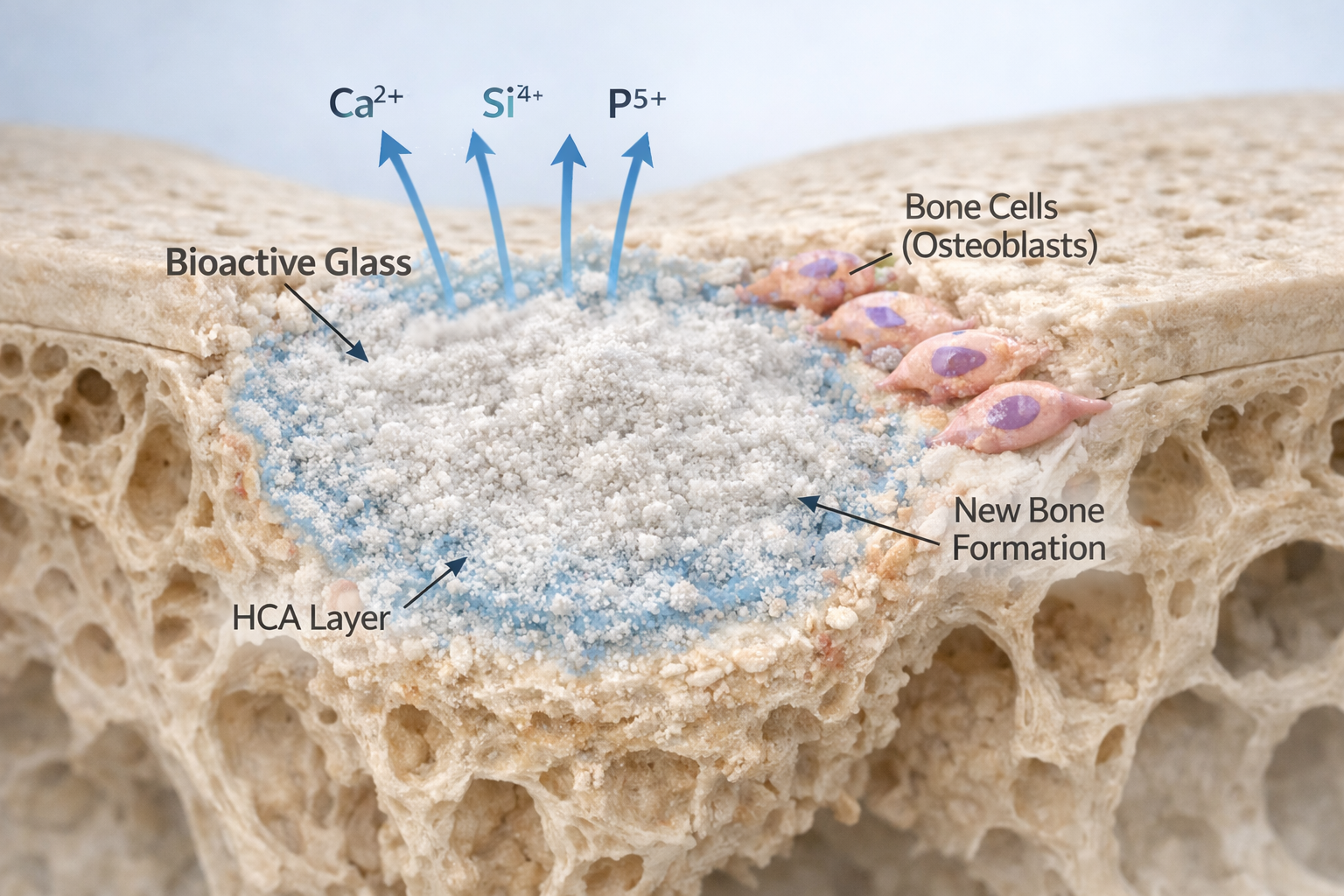
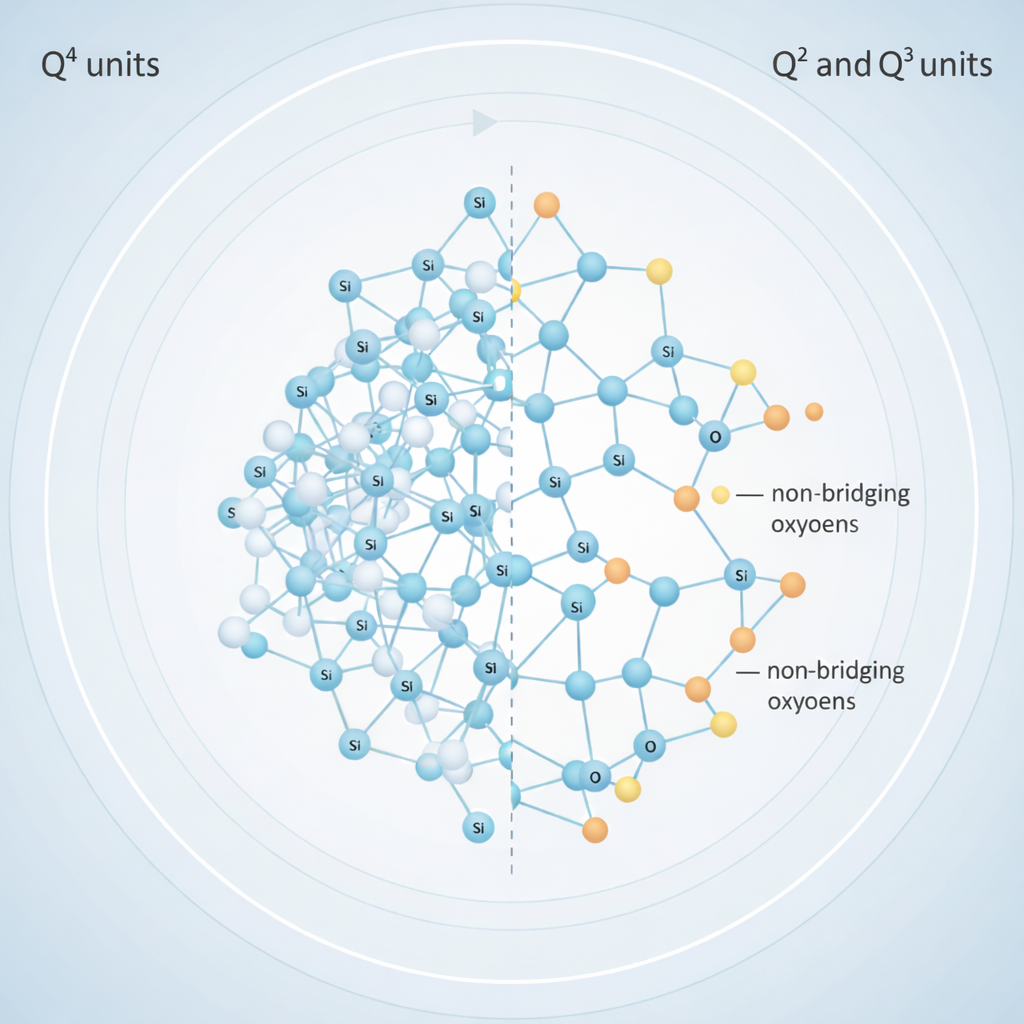
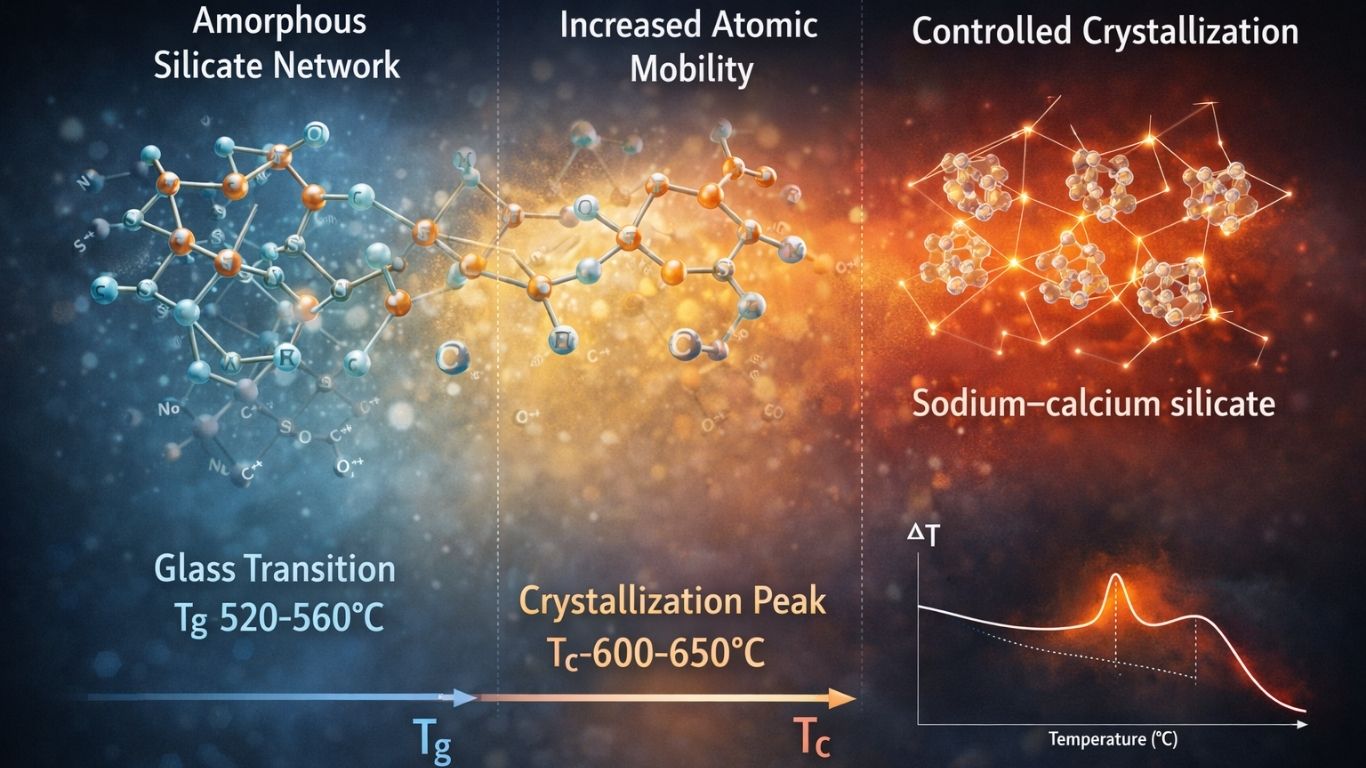
Bioactive glass is a silicate-based biomaterial known for its exceptional bioactivity and biocompatibility. When in contact with body fluids, it forms a hydroxycarbonate apatite (HCA) layer — similar to natural bone mineral — and releases therapeutic ions that activate genetic and biochemical responses in nearby cells.
These bioactive ions not only support bone tissue growth but also influence stem cells by:
- Stimulating cell proliferation
- Inducing osteogenic differentiation
- Promoting extracellular matrix mineralization
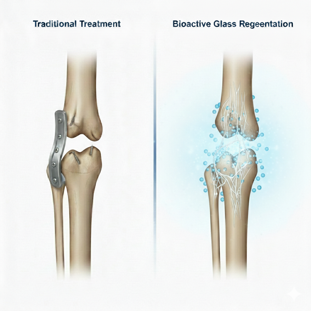
Such features make bioactive glass a cornerstone material in bone grafts, scaffolds, coatings, and regenerative implants.
However, the real measure of its success lies in how effectively it can drive stem cells toward osteogenesis — a process quantified using biochemical assays like ALP and histochemical stains like Alizarin Red S.
Why the ALP Assay Matters in Bioactive Glass Research
What Is ALP and Why It’s Important
Alkaline phosphatase (ALP) is an enzyme predominantly expressed during the early stages of osteoblast differentiation. It plays a key role in hydrolyzing phosphate esters, thereby increasing the local phosphate concentration needed for hydroxyapatite formation, the mineral backbone of bone tissue.
High ALP activity reflects that stem cells are transitioning into osteoblasts, marking the onset of bone formation. For researchers working with bioactive glass, measuring ALP activity is therefore one of the most reliable and early indicators of osteogenic differentiation.
Stem Cell Response to Bioactive Glass
Various types of stem cells—such as mesenchymal stem cells (MSCs), adipose-derived stem cells (ASCs), and dental pulp stem cells—have been shown to respond positively to bioactive glass exposure. Studies consistently demonstrate that when cultured with bioactive glass or its ion-rich extracts, these cells exhibit significantly elevated ALP levels, proving that bioactive glass accelerates their osteogenic commitment.
Research by Wang et al. (2018) revealed that human adipose stem cells exposed to bioactive glass displayed markedly increased ALP activity and enhanced bone-related gene expression. Similar findings were observed by Abdelaziz et al. (2024), confirming that bioactive glass nanoparticles promote both proliferation and differentiation of dental stem cells.
Conducting the ALP Assay with Bioactive Glass
The ALP assay helps quantify the enzymatic activity associated with early bone formation. Here’s how it’s typically performed in bioactive glass studies:
1. Cell Culture and Differentiation
- Stem cell seeding: Cells are plated at optimal density in tissue culture wells.
- Exposure to bioactive glass: Cells are grown either directly on bioactive glass scaffolds or particles, or in media conditioned with bioactive glass extracts.
- Osteogenic induction: A specialized medium containing dexamethasone, ascorbic acid, and β-glycerophosphate is added to encourage bone differentiation.
- Incubation: The cultures are maintained for 7–21 days, with regular media changes to replenish nutrients and ions released by the bioactive glass.
2. Measuring ALP Activity
- At predetermined intervals (7, 14, and 21 days), cells are lysed to extract intracellular enzymes.
- A substrate such as p-nitrophenyl phosphate (pNPP) is added. ALP converts this substrate into a yellow-colored product, measurable via spectrophotometer at 405 nm.
- Fluorescent alternatives like ELF-97 can be used for more sensitive detection.
- To ensure accuracy, ALP activity is normalized to total protein or DNA content, accounting for differences in cell proliferation.
3. Data Interpretation
An increase in ALP activity in bioactive glass-treated samples indicates that the material effectively stimulates early osteogenic differentiation.
This biochemical data is often validated with:
- Gene expression analysis (RUNX2, Osteocalcin, Osteopontin)
- Mineralization assays such as Alizarin Red S staining for late-stage confirmation.
Alizarin Red S (ARS) Staining: Evaluating Late-Stage Differentiation with Bioactive Glass
Purpose and Mechanism
While the ALP assay highlights early differentiation, Alizarin Red S (ARS) staining focuses on mineralization, the final step of osteogenic differentiation. It is a histochemical technique that detects calcium deposits formed by mature osteoblasts.
The ARS dye has a strong affinity for calcium ions, particularly those in hydroxyapatite crystals — the same mineral structure found in bone. When applied to cultured cells, ARS binds to these deposits, producing vivid red-orange coloration. The intensity of the color corresponds to the amount of mineralization, allowing both qualitative visualization and quantitative measurement.
Role of Bioactive Glass in ARS Staining
Bioactive glass enhances mineralization by releasing bioactive ions such as calcium and silicon, which are key stimulators of osteogenic gene expression and extracellular matrix formation.
When stem cells are cultured with bioactive glass scaffolds or conditioned media, they show:
- Higher calcium deposition,
- More intense ARS staining, and
- Stronger mineral nodule formation under microscopic analysis.
These findings prove that bioactive glass not only triggers osteogenic differentiation but also supports complete mineralized matrix formation, confirming its osteoconductive and osteoinductive properties.
Detailed ARS Protocol for Bioactive Glass Studies
- Culture Phase:
Stem cells are grown with bioactive glass materials in osteogenic induction medium for 21–28 days to allow sufficient mineralization. - Fixation:
Cells are fixed with 4% paraformaldehyde to preserve the matrix and mineral deposits. - Staining:
The fixed cells are incubated with 1–2% Alizarin Red S solution (pH ~4.2) for about 30 minutes. The dye binds calcium-rich regions, producing bright red-orange coloration. - Washing and Imaging:
Excess dye is washed off with distilled water, and stained cultures are observed under light microscopy for qualitative assessment. - Quantification:
For quantitative analysis, the dye bound to calcium is eluted using cetylpyridinium chloride, and its absorbance is measured at 562–570 nm using a spectrophotometer.
This data provides a precise measure of mineralization, allowing researchers to compare the effects of different bioactive glass formulations, ion compositions, and exposure durations.
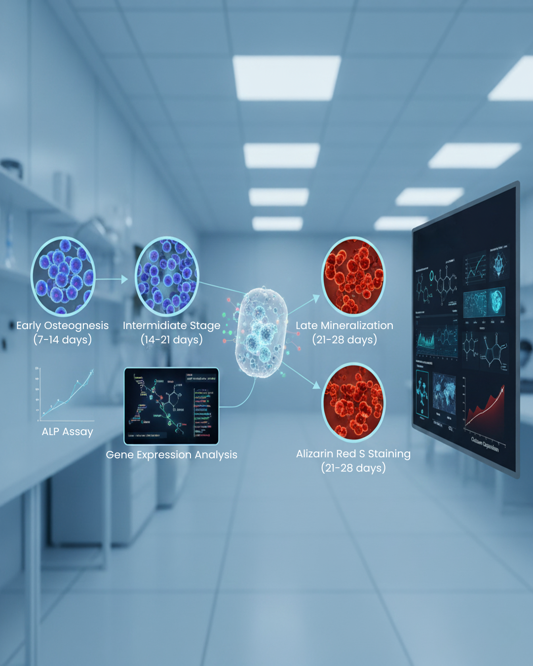
Correlating ALP and ARS Findings in Bioactive Glass Research
In bone tissue engineering research, ALP and ARS results are often evaluated together to provide a complete picture of bioactive glass performance:
| Stage of Differentiation | Marker/Assay Used | Observation in Bioactive Glass Studies |
| Early Stage (7–14 days) | ALP Assay | High ALP enzyme activity indicates early osteogenic commitment. |
| Intermediate Stage (14–21 days) | Gene Expression (RUNX2, Osteopontin) | Upregulation of bone-related genes shows active differentiation. |
| Late Stage (21–28 days) | Alizarin Red S Staining | Strong red coloration indicates calcium-rich mineralized matrix formation. |
Together, these techniques confirm that bioactive glass plays a vital role in guiding stem cells through all stages of osteogenic differentiation — from commitment to complete mineralization.
Scientific Validation of Bioactive Glass in Osteogenesis
Several landmark studies have established the correlation between bioactive glass and osteogenic differentiation:
- Wang et al. (2018) demonstrated that human adipose-derived stem cells exposed to bioactive glass exhibited enhanced osteogenic gene expression and ALP activity, driven by MAPK signaling pathways.
- Reilly et al. (2007) compared rat and human bone marrow stem cell responses to 45S5 bioactive glass, confirming species-specific differences but consistent osteogenic stimulation.
- Chen et al. (2006) developed 45S5 Bioglass® scaffolds for bone tissue engineering, highlighting superior mineralization and biocompatibility.
- Bernar et al. (2022) optimized Alizarin Red S assays to improve detection sensitivity of mineralization in osteoblast cultures — crucial for evaluating bioactive glass-induced bone formation.
These findings collectively affirm that bioactive glass is more than a passive scaffold—it is an active participant in stem cell regulation and bone regeneration.
Conclusion: Bioactive Glass as a Catalyst for Bone Regeneration
The integration of stem cell technology and bioactive glass biomaterials represents a groundbreaking shift in regenerative medicine. Through methods like ALP assays and Alizarin Red S staining, scientists can clearly observe and quantify how bioactive glass drives osteogenic differentiation and mineralization.
With its ability to release biologically active ions, bioactive glass functions as both a structural framework and a signaling platform, making it a superior choice for next-generation bone graft substitutes, coatings, and scaffolds.
As research advances, the continued optimization of staining methods, assay sensitivity, and glass compositions will further unlock the regenerative potential of bioactive glass, shaping the future of bone tissue engineering and clinical applications.
Contact us through Synthera Biomedical social platforms to stay informed about pioneering bioactive glass research and clinical applications. Follow us on Instagram for product launches and research updates. Join the conversation on Facebook to access valuable resources and community news.