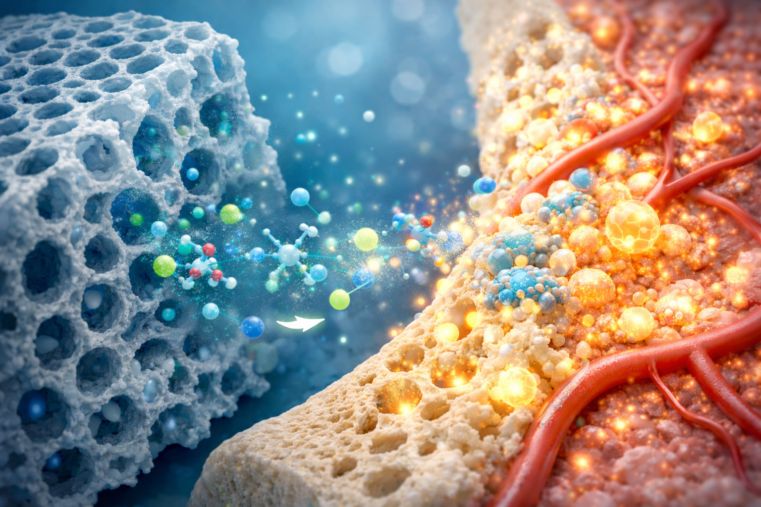

All of the complexities in the body during in vivo tests cannot be captured by in vitro methods. However, they come before in vivo tests because they are useful predictors of how recently created biomaterials, like Bioactive glass, might behave when they come into contact with tissue in vivo. The fact that biocompatibility is a property of the entire system under study, not just a biomaterial, should not be overlooked. We refer to this as cytocompatibility, i.e. compatibility between a specific cell type and a biomaterial in vitro. Either directly (contact test), indirectly (agar or filter), or through the dissolution products released into the culture medium (elution test), cells come into contact with the Bioactive glass. A Bioactive glass’s possible cytotoxicity is assessed before its mode of action is investigated. The ISO 10993-5 standard for biological safety evaluation (Biological evaluation of medical devices – Tests for in vitro cytotoxicity) outlines standardized tests.
Spectrophotometry, fluorimetry, and microscopy are examples of experimental techniques that can be used to assess a variety of parameters, such as metabolic activity, cell membrane integrity, morphology, and proliferation. Microscopy-based techniques can provide information on cell morphology and, especially in electron microscopy studies, on how cells interact with the surface of materials, even though quantitative techniques like spectrophotometric techniques are preferred. The results of these tests, which are covered in greater detail in the chapter on “In vitro cytotoxicity tests,” provide details about a material’s cytocompatibility, i.e. e. whether or not it has any negative effects on cultured cells. Liu et al. recently reviewed the procedures used for medical device safety testing generally. An OECD (Organization for Economic Co-operation and Development) document also lists the viability testing techniques for cell cultures along with their benefits and drawbacks. (1)
Cell Model:
Choosing the right kind of cells is the first query. For in vitro testing, a variety of cell types are employed; the selection is based on the assay undertaken as well as the laboratory’s amenities. For the tests, primary cells and cell lines—including cells from various tissues and organisms (human or animal cells)—are utilized. The majority of bone cells and other mammalian cells are adherent. (2)
Primary cells are extracted straight from the living thing. Examples include human osteoblasts that were separated from animal samples, usually from rats or mice, or from trabecular bone fragments taken during surgery or biopsies. Although they are usually more realistic models, their results are not always reproducible because they can differ depending on the donor as well as the culture conditions. In contrast, commercially available immortalized cells are known as continuous cell lines. Their results may be less physiologically relevant, but they are easier to obtain and more reproducible. But for simple cytotoxicity testing, cell lines are adequate, and using human cells in the initial testing phase is also not required. (3)
Specialized cells are needed for additional testing of Bioactive glass for bone regeneration. i.e. Depending on the intended use, specific cell lines—primary bone cells or multipotent cells—can be selected. Since Bioactive glass is primarily used in bone regeneration, the cell lines used in more complex tests are typically derived from bone tissue and are known as “osteoblast like cells.
Cytotoxicity and other in vitro cell culture test results are typically cell type dependent. Furthermore, different cell types usually require different cell culture growth medium compositions. This variation in composition can significantly affect Bioactive glass ion release and apatite mineralization, which may in turn affect the results of any Bioactive glass in vitro cell culture studies. (4)
In vitro cytotoxicity tests
Cytotoxicity tests are used to determine the potential negative effects of materials and/or their extracts on cells. Cell viability is the ability of a cell in culture to maintain normal shape and function. It is characterized by morphology, cell membrane integrity, or metabolic activity. Spectrophotometry, fluorimetry or luminometry, usually combined with chromogenic, fluorogenic or light-emitting substrates, can be particularly useful for quantitative evaluation, and it is usually based on chromogenic, fluorogenic or light-emitting substrates, respectively.
The form and appearance of cells is known as cell morphology (e.g. G. polygonal or spindle-shaped), and it depends on the type of cell. Adherent cells that are in good health adhere to the surface and proliferate. A rounded shape and separation from the surface are the primary indicators of cell deterioration. Cytoskeletal architecture is necessary for maintaining both cell adhesin and cell shape. As a result, fluorescence labeling and visualization of cytoskeleton components primarily microfilaments made of f-actin are common. DNA binding dyes are typically used to counterstain cell nuclei. In addition to fluorescence microscopy, optical or scanning electron microscopy are used to assess cell morphology. (5,6)
Another frequently measured parameter is cell proliferation. Cells divide through a series of events known as the cell cycle. The actual division of the cell into two daughters during mitosis and DNA synthesis, which takes place during the S-phase, are the two main events in the cell cycle. Nonetheless, there is no requirement that cell viability and proliferation be correlated. Cytostatic substances are those that only prevent cell division rather than killing cells. Cell proliferation can be described as the actual percentage of proliferating cells in the culture or as a gradual increase in the number of cells.
References:
- Bellucci D, Veronesi E, Dominici M, Cannillo V. On the in Vitro Biocompatibility Testing of Bioactive Glasses. Materials (Basel). 2020 Apr 12;13(8):1816.
- Chen Q.Z.Z., Thompson I.D., Boccaccini A.R. 45S5 Bioglass-derived glass-ceramic scaffolds for bone tissue engineering. Biomaterials. 2011;27:2414–2425.
- Bellucci D., Veronesi E., Strusi V., Petrachi T., Murgia A., Mastrolia I., Dominici M., Cannillo V. Human mesenchymal stem cell combined with a strontium enriched bioactive glass: An ex-vivo model for bone regeneration. Materials. 2019;12:3633.
- Tsigkou O, Jones JR, Polak JM, Stevens MM. Differentiation of fetal osteoblasts and formation of mineralized bone nodules by 45S5 Bioglass conditioned medium in the absence of osteogenic supplements. Biomaterials. 2009;30:3542–3550.
- Hoppe A., Guldal N.S., Boccaccini A.R. A review of the biological response to ionic dissolution products from bioactive glasses and glass-ceramics. Biomaterials. 2011;32:2757–2774.
- Rismanchian M, Khodaeian N, Bahramian L, Fathi M, Sadeghi-Aliabadi H. In-vitro Comparison of Cytotoxicity of Two Bioactive Glasses in Micropowder and Nanopowder forms. Iran J Pharm Res. 2013.